Prenatal Ultrasound: Essential Tool For Fetal Health And Development Evaluation

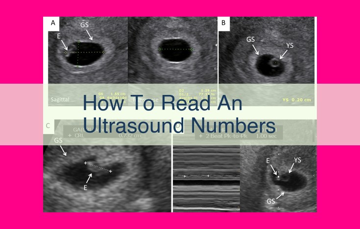
Ultrasound numbers, assessed during prenatal ultrasounds, provide valuable information about fetal growth and development. Key measurements include BPD (biparietal diameter), CRL (crown-rump length), and FL (femur length), which help determine gestational age. Ultrasounds also detect fetal anomalies and conditions like Down Syndrome, allowing for early intervention. In multiple pregnancies, ultrasound monitors fetal growth and assesses potential complications. Accurate interpretation by sonographers and OB-GYNs is crucial for optimal pregnancy care, providing reassurance, aiding decision-making, and enhancing fetal well-being.
Ultrasonography: A Vital Tool in Pregnancy Care
In the realm of pregnancy, ultrasonography holds immense significance as a non-invasive imaging technique that illuminates the world within the womb. Through the emission of high-frequency sound waves, an ultrasound paints a vivid picture of your developing baby, providing a wealth of information about its growth, well-being, and any potential concerns.
Ultrasonography serves as an invaluable tool for healthcare professionals, allowing them to monitor the baby’s growth, assess its anatomical structures, and identify potential abnormalities that may require further evaluation or treatment. It plays a crucial role in ensuring the health and well-being of both the mother and the developing fetus, empowering them to make informed decisions throughout the pregnancy journey.
Key Structures and Measurements in Pregnancy Ultrasounds
During an ultrasound examination in pregnancy, skilled sonographers meticulously assess a range of key structures and meticulously take measurements to glean vital insights into the health and well-being of both the developing baby and the expecting mother. These crucial structures and measurements provide a comprehensive picture of the baby’s growth and development, allowing healthcare professionals to monitor the pregnancy closely.
Measuring the Fetus: A Window into Growth and Gestational Age
One of the primary focuses of an ultrasound examination is assessing the fetus’s growth. Sonographers measure various parameters to determine the baby’s gestational age and overall development. These measurements include:
- BPD (Biparietal Diameter): This measures the distance between the two sides of the fetal head, helping to estimate the baby’s size and maturity.
- CRL (Crown-Rump Length): This measurement extends from the top of the baby’s head to the rump (buttocks). It is commonly used to determine gestational age in the early stages of pregnancy.
- FL (Femur Length): This measurement assesses the length of the baby’s thigh bone, providing an indication of their overall skeletal development.
Monitoring Fetal Health: Beyond Growth
Beyond determining gestational age, ultrasound examinations also evaluate the fetus’s anatomical structures and organ systems for any potential anomalies or concerns. These assessments include:
- Placenta: The placenta, a vital organ supporting the baby’s development, is assessed for its position, size, and overall appearance.
- Amniotic Fluid: The amount and clarity of the amniotic fluid surrounding the baby are evaluated as well.
- Doppler Imaging: This advanced ultrasound technique allows healthcare professionals to visualize and assess blood flow in the fetus’s heart and other blood vessels, providing insights into their circulatory system’s functionality.
Fetal Anomalies and Conditions Detectable through Ultrasonography
Ultrasound examinations during pregnancy play a crucial role in identifying various fetal anomalies and conditions. These include:
Anencephaly
Anencephaly is a severe neural tube defect where the baby’s brain does not develop properly. Ultrasound can detect this condition as early as the first trimester, allowing for informed decision-making and potential prenatal care options.
Down Syndrome
Down Syndrome is a genetic condition that causes intellectual disability and physical features. Ultrasound can screen for Down Syndrome by measuring the thickness of the baby’s neck fold and looking for other markers. Further testing may be recommended if abnormalities are detected.
Preterm Birth
Preterm birth, occurring before 37 weeks of gestation, can lead to health complications for the baby. Ultrasound can assess the length of the baby’s cervix, which can help determine the risk of preterm labor and guide appropriate interventions.
Significance of Early Detection
Early detection of fetal anomalies and conditions through ultrasound is incredibly important. It allows for prenatal counseling, where families can make informed decisions about their pregnancy, consider potential treatments or interventions, and prepare for the future needs of their child.
Potential Interventions
Depending on the specific anomaly or condition diagnosed, various interventions may be available. For example, some neural tube defects can be surgically corrected during pregnancy to improve the baby’s outcomes. In other cases, medications or therapies may be recommended to manage the effects of the condition.
Multiple and High-Risk Pregnancies: The Role of Ultrasound
When it comes to high-risk pregnancies, ultrasound takes on an even more critical role. Women with pre-existing conditions, such as diabetes or heart disease, face increased risks during pregnancy, and ultrasound allows doctors to monitor both the mother and the baby closely. Regular ultrasounds can detect potential complications, such as placental abruption or preeclampsia, allowing for early intervention and improved outcomes.
Ultrasound also plays a unique role in pregnancies with multiples. Monitoring the growth and development of twins or triplets can be more challenging, but ultrasound helps doctors evaluate each baby’s anatomy, ensure they are growing properly, and identify potential complications. It’s also crucial for determining the type of multiples (e.g., identical or fraternal), as this can impact pregnancy management.
During an ultrasound for multiples, the doctor may measure the nuchal translucency (the fluid-filled space at the back of the baby’s neck) and perform Doppler studies (which use sound waves to assess blood flow) to assess the baby’s health. By monitoring the babies’ growth, position, and any potential complications, ultrasound helps doctors make informed decisions and optimize pregnancy care.
Interpreting Ultrasound Results: A Collaborative Approach
In the world of pregnancy, ultrasound plays a pivotal role in assessing fetal well-being and monitoring the health of both the mother and child. While these images provide invaluable insights, interpreting ultrasound results accurately is crucial to ensure optimal care.
This delicate task involves a collaborative effort among highly skilled professionals:
Sonographers: The Eyes Behind the Images
Sonographers, the individuals operating the ultrasound equipment, possess specialized training in capturing high-quality images. Their keen eye for detail enables them to identify and measure key anatomical structures, such as the fetus, uterus, and placenta. These measurements serve as essential indicators of gestational age and fetal growth.
OB-GYNs: Medical Expertise and Interpretation
Obstetricians and gynecologists (OB-GYNs) bring their medical knowledge and expertise to interpret the ultrasound findings. They assess the baby’s heart rate, growth patterns, anatomy, and any potential abnormalities. Based on their observations, OB-GYNs provide guidance on necessary follow-up care, including additional testing or interventions.
Genetic Counselors: Providing Clarity and Support
In cases where ultrasound results suggest a potential fetal anomaly, genetic counselors play a vital role. They provide compassionate guidance and information about genetic conditions, their implications, and available options. Genetic counselors help expectant parents navigate complex medical decisions, empowering them to make informed choices about their pregnancy.
Accurate Interpretation: The Cornerstone of Informed Care
Accurate interpretation of ultrasound results is paramount for optimal pregnancy care. It allows healthcare providers to:
- Confirm gestational age and track fetal growth
- Identify potential complications, such as preterm birth or pregnancy-related hypertension
- Detect fetal anomalies, enabling early intervention and support
- Provide reassurance and peace of mind to expectant parents
The Importance of Collaboration and Communication
Effective communication and collaboration among sonographers, OB-GYNs, and genetic counselors ensure consistent and precise interpretation of ultrasound results. Regular discussions, consultations, and case reviews foster a shared understanding of fetal health and guide appropriate decision-making. This collaborative approach contributes to the well-being of both the mother and child throughout the pregnancy journey.
Benefits and Limitations of Ultrasonography
Ultrasound has revolutionized pregnancy care, providing expectant mothers and healthcare providers with unparalleled insights into the developing fetus. While its benefits are undeniable, it’s essential to acknowledge its limitations to ensure informed decision-making.
Benefits of Ultrasonography:
-
Early Detection: Ultrasound allows for the early detection of a wide range of fetal anomalies and conditions. This enables timely intervention and treatment, significantly improving outcomes for both mother and baby.
-
Reassurance: For many expectant mothers, ultrasound provides a much-needed sense of reassurance. Seeing their unborn child on screen can dispel anxiety and instill a sense of connection with their developing little one.
-
Planning for Delivery: Accurate ultrasound measurements help determine gestational age, which is crucial for _planning delivery_. This information ensures appropriate timing and preparation for labor, improving the chances of a safe and successful birth.
Limitations of Ultrasonography:
-
False Negatives and False Positives: While ultrasound is a powerful tool, it’s not foolproof. There’s a potential for false negatives, meaning the scan may miss certain anomalies. Similarly, false positives can lead to unnecessary anxiety and follow-up procedures.
-
Operator Dependency: The accuracy of ultrasound results heavily depends on the skill and experience of the sonographer. Variations in technique and interpretation can lead to discrepancies in findings.
-
Certain Conditions May Require Further Tests: Some conditions, such as fetal heart defects, may not be fully visualized on ultrasound. In such cases, additional tests or procedures, like echocardiography, may be necessary for a more comprehensive assessment.
Understanding the limitations of ultrasound is crucial for expectant mothers. While it provides invaluable information, it’s not a substitute for regular prenatal care, genetic counseling, and other diagnostic tests. By striking the right balance between ultrasound and other modalities, healthcare providers can ensure a comprehensive and accurate picture of the developing fetus, empowering expectant mothers to make informed choices throughout their pregnancy journey.