Combat Chest Pain From Posture: A Holistic Guide To Muscle Imbalances &Amp; Heart Health Optimization

To alleviate chest pain caused by poor posture, focus on addressing the underlying musculoskeletal imbalances. Engage in posture-correcting exercises to strengthen the trapezius, pectoralis minor, and other affected muscles. Simultaneously, release tension in tight muscles like the levator scapulae and scalenes. Additionally, ensure cardiovascular health by managing coronary artery disease and optimizing blood flow to the heart.
Muscles Involved in the Pain Pathway: A Comprehensive Exploration
Muscles: The Unsung Heroes of Pain Perception
In the intricate tapestry of pain, muscles play a pivotal role, orchestrating the symphony of pain signals that travel throughout the body. Muscles are not mere passive players; they are active participants in the generation and transmission of pain. Understanding their involvement is crucial for unraveling the complex mechanisms of pain.
Trapezius: The Neck’s Guardian, Now Pain’s Herald
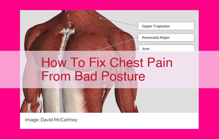
The broad trapezius muscle, spanning the length of the neck, can be a harbinger of pain when overexerted. Its relentless contraction can strain the neck, leading to stiffness, tenderness, and headaches. When pain grips the trapezius, every turn of the head becomes a symphony of discomfort.
Pectoralis Minor: The Chest’s Enigma, Unmasking Pain’s Secrets
Beneath the pectoralis major lies the enigmatic pectoralis minor. This smaller muscle, when tight or strained, can trigger referred pain in the chest and shoulder. It lurks in the shadows, its presence often overlooked until pain reveals its hidden affliction.
Rhomboid: The Shoulder’s Stabilizer, Now a Source of Woe
The rhomboid muscles, nestled between the shoulder blades, are responsible for stabilizing the shoulders. However, when they become overexerted or injured, they can revolt, unleashing pain that radiates through the shoulders and neck. The once-reliable rhomboid now becomes a relentless source of discomfort.
Latissimus Dorsi: The Back’s Powerhouse, Now a Pawn of Pain
The latissimus dorsi, the largest muscle of the back, is a force to be reckoned with. But when it succumbs to strain or injury, it can unleash a torrent of pain that ripples down the back and into the arms. The latissimus dorsi, once a pillar of strength, now buckles under pain’s unrelenting grip.
Levator Scapulae: The Neck’s Lifter, Now a Pain Inflictor
The levator scapulae muscle, connecting the neck to the shoulder blade, has a crucial role in lifting the shoulder. But when it’s overworked or injured, it becomes a pain generator, radiating discomfort through the neck and shoulders. The once-essential levator scapulae now turns into a relentless source of agony.
Scalene Muscles: The Neck’s Guardians, Now Pain’s Accomplices
The scalene muscles, located on the sides of the neck, are responsible for rotating and flexing the neck. However, they can also become harbingers of pain when they become tight or strained. The scalene muscles, once protectors of the neck, now become its tormentors, inflicting discomfort that radiates through the head and shoulders.
Muscles Involved in the Pain Pathway
As muscles contract and relax, they can become strained or injured, leading to the activation of nociceptors – specialized nerve endings that detect pain. Several muscles in the upper body play significant roles in pain generation and transmission:
-
Trapezius: This large, triangular muscle extends from the base of the skull to the mid-back. It helps stabilize the shoulder and move the head and neck. When strained, it can cause pain in the neck, shoulders, and upper back.
-
Pectoralis Minor: This small muscle connects the breastbone to the shoulder blade. It helps pull the shoulder forward and rotate the shoulder joint. Tightness or strain in the pectoralis minor can result in pain in the chest and front of the shoulder.
-
Rhomboid: A group of muscles located between the shoulder blades, the rhomboids help retract (pull back) the shoulder blades. Poor posture or overuse can strain these muscles, causing pain in the upper back and between the shoulder blades.
-
Latissimus Dorsi: The largest muscle in the back, the latissimus dorsi extends from the mid-back to the hip. It helps with pulling, rotating, and adducting (bringing the arm towards the body) the arm. Strain or injury to the latissimus dorsi can lead to pain in the back, shoulder, or armpit.
-
Levator Scapulae: This muscle runs from the base of the skull to the shoulder blade and assists in elevating the shoulder. Tightness or strain in the levator scapulae can cause pain in the neck, shoulder, and upper back.
-
Scalene Muscles: Three scalene muscles (anterior, middle, and posterior) are located on the sides of the neck. They help bend the neck laterally (to the side) and rotate the head. Overuse or poor posture can strain these muscles, resulting in pain in the neck, shoulder, and head.
Coronary Arteries and Pain: The Story of Angina and Heart Attack
In the intricate tapestry of our body, an unseen battle wages when our coronary arteries, the vital vessels that nourish the heart, become compromised. This conflict manifests as chest pain, a harbinger of underlying cardiac distress.
Angina Pectoris: A Cry for Oxygen
Angina pectoris, the medical term for chest pain, is a warning that your heart is struggling for oxygen. It occurs when the blood flow through the coronary arteries is partially blocked, usually by fatty deposits called plaques. As the heart works harder to pump blood against this obstruction, it demands more oxygen, which cannot be met due to the narrowed arteries. This mismatch between demand and supply triggers angina pain.
The pain of angina is often described as a crushing, squeezing, or burning sensation, located in the center of the chest. It may radiate to the left arm, neck, jaw, or back. Angina pain typically lasts for a few minutes and is relieved by rest or medications that widen the coronary arteries.
Myocardial Infarction: A Heart Attack
When the blockage in a coronary artery becomes complete, severing blood flow to a portion of the heart, a myocardial infarction, commonly known as a heart attack, occurs. The deprived heart tissue begins to die within minutes, causing severe and prolonged chest pain.
The pain of a heart attack is often described as a stabbing, tearing, or aching sensation, along with a feeling of pressure or tightness. It typically lasts for more than 20 minutes and is not relieved by rest. Other symptoms may include shortness of breath, nausea, vomiting, and sweating.
Understanding the Pathophysiology
The connection between coronary artery disease and chest pain lies in the heart’s dependence on a constant supply of oxygenated blood. When plaque builds up in the arteries, it narrows the passageway, reducing blood flow and oxygen delivery. This triggers a cascade of events that leads to pain.
In angina, the reduced blood flow is enough to cause temporary oxygen deprivation, resulting in chest pain. However, if the blockage is complete, as in a heart attack, the heart tissue is permanently damaged, leading to severe pain and potential life-threatening consequences.
Recognizing and Responding to Chest Pain
Chest pain should never be ignored, especially if it is accompanied by other symptoms of angina or a heart attack. Prompt medical attention is crucial to diagnose the underlying cause and receive appropriate treatment.
If you experience chest pain, stay calm and seek medical help immediately. Aspirin can help reduce the risk of a heart attack if taken promptly, but do not take it without consulting a medical professional.
Coronary Arteries and Chest Pain
Unlocking the Pain Pathways
Our bodies are intricate networks of interconnected systems, each with its own unique role to play. When it comes to pain, muscles, nerves, and cardiovascular structures dance together in a complex symphony of communication. Let’s delve into the pain pathway involving these entities, starting with the cardiovascular connection.
The Heart of the Matter: Coronary Arteries and Chest Pain
The coronary arteries are the vital blood vessels that supply oxygen-rich blood to the heart. When these arteries become narrowed or blocked by plaque buildup, it can restrict blood flow, triggering a cascade of events that lead to chest pain.
Angina Pectoris: A Wake-Up Call
Angina pectoris is a type of chest pain that occurs when the heart doesn’t receive enough oxygen. It’s characterized by a tightness, pressure, or squeezing sensation in the chest, often accompanied by shortness of breath, nausea, and sweating. Angina pectoris is a warning sign that you may be at risk of a more severe heart event.
Myocardial Infarction: A Silent Killer
If the blockage in a coronary artery becomes complete, it can lead to a myocardial infarction, commonly known as a heart attack. When part of the heart muscle is deprived of oxygen for an extended period, it can result in permanent damage or even death. Myocardial infarctions often present with sudden, severe chest pain that lasts for more than 20 minutes. Other symptoms may include pain radiating to the arms, neck, or back, sweating, shortness of breath, and nausea.
Seeking Help: The Importance of Prompt Action
If you experience chest pain, seek medical attention immediately. Early diagnosis and treatment can significantly improve outcomes and prevent serious complications. Remember, chest pain is a vital signal from your body, one that should never be ignored. By understanding the connection between coronary artery disease and chest pain, you empower yourself to take proactive steps towards maintaining a healthy heart.
Subheading: Cervical and Thoracic Nerves in Pain Transmission
In the intricate dance of pain, the central nervous system (CNS) acts as the conductor, orchestrating every sensation we experience. Pain, a complex and multifaceted journey, begins with the initial nociceptive impulse, which is then transmitted through a network of nerves to the spinal cord and ultimately to the brain for processing.
Cervical and thoracic nerves play a crucial role in this pain transmission pathway, acting as the messengers that convey these signals from the periphery to the CNS. These nerves traverse the neck and chest regions, branching out to innervate various muscles, joints, and organs.
The cervical nerves, originating from the spinal cord in the neck, are responsible for sensory and motor function in the head, neck, and upper limbs. Among these nerves, the C4-C8 nerve roots are particularly significant in pain transmission. They innervate the muscles and joints of the neck, shoulders, upper back, and arms. Any irritation or damage to these nerves can result in neck pain, shoulder pain, and radiating pain along the arm.
Thoracic nerves, emerging from the spinal cord in the chest, extend to the chest wall, abdomen, and upper extremities. The T2-T6 nerve roots are key players in transmitting pain signals from the chest and upper abdomen. They innervate the muscles and joints of the chest, back, and rib cage. Pain in these regions, such as chest pain or back pain, can often be attributed to issues with these thoracic nerves.
The intricate network of cervical and thoracic nerves forms a gateway through which pain signals flow. Understanding their anatomy and function is essential for deciphering the origins of pain and guiding appropriate treatment strategies.
Cervical and Thoracic Nerves: Gatekeepers of Pain Transmission
In the intricate tapestry of our body’s pain pathway, cervical and thoracic nerves serve as crucial conduits, relaying distress signals from our muscles and organs to the central command center of our brain. These nerves play a fundamental role in our perception and management of pain.
Anatomy and Function:
The cervical nerves (C1-C8) emerge from the spinal cord in our neck, while the thoracic nerves (T1-T12) originate from the spinal cord in our chest. These nerves branch out like intricate threads, innervating the muscles, skin, and organs in their respective regions. Each nerve has specialized sensory and motor fibers, allowing us to sense touch, temperature, and pain, as well as control muscle movement.
Pain Transmission:
When pain arises from musculoskeletal structures, nociceptors (specialized pain receptors) in the muscles activate. These nociceptors send signals along the sensory fibers of cervical or thoracic nerves to the spinal cord. The spinal cord then relays these signals to the brain, where they are interpreted as pain.
The transmission of pain signals is not a straightforward process. Along the way, gate control mechanisms within the spinal cord can modulate the intensity of pain. These mechanisms act like a filter, allowing only certain signals to pass through.
Specific Roles:
- Cervical Nerves (C3-C5): These nerves convey pain signals from the muscles and skin in the neck, shoulder, and upper back. They are particularly involved in headaches, neck pain, and pain radiating down the arms (known as radiculopathy).
- Thoracic Nerves (T1-T5): These nerves receive pain signals from the chest wall, lungs, and heart. They play a critical role in conditions such as chest pain, angina, and myocardial infarction (heart attack).
Understanding the anatomy and function of cervical and thoracic nerves is essential for effectively managing pain. By targeting these nerves through therapies such as nerve blocks or medications, we can disrupt the pain pathway and provide much-needed relief.